ISSN: 2688-562X
Review Article
Spinal Arachnoid Cysts: Clinical and Technical Study
Kamel Bahri1, Mohamed Chabaane1, Ghassen Gader1*, Sofiene Bouali2, Hafedh Jemel2 and Ihsèn Zammel1
- 1Department of Neurosurgery, Trauma and Burns Center, Ben Arous, Tunisia
- 2Department of Neurosurgery, National Institute of Neurology, Tunis, Tunisia
- *Corresponding author: Ghassen Gader, Department of Neurosurgery, Trauma and Burns Center, Ben Arous, Tunisia
- Received: Dec 22, 2018 Accepted: Jan 18, 2019 Published: Jan 24, 2019
- Keywords: Colorectal Cancer; Genetics; Genes
Introduction
Spinal arachnoid cysts are rarelesions that account for nearly 1 % of all spinal lesions [1]. Despite the fact that these lesions are most generally asymptomatic, they may lead to neurologic manifestations by compression of the medulla, the cauda equine or nerve roots [2]. Spinal arachnoid cysts are surgically curable. Nevertheless, they have not been yet subject of deep studies, as most of the publications are single case reports. Through this paper, we report our experience in the field of treatment of spinal arachnoid cysts through the study of 13 patients.
Methods
We report a retrospective study of 13 patients managed between 2010 and 2017 for a spinal arachnoid cyst. Post traumatic arachnoid cysts were not included in our study. All our patients had a spinal MRI. 3 of them had also a computed myelography. 12 patients underwent surgery through various approaches including cyst excision, cyst shunt or cyst punctures.
Results
(Table 1) The mean age for our patients was 36 years, with extremes ranging between 4 and 69 years old. 10 of our patients were female gendered with a sex ratio female/male at 3, 33/1. All our patients were symptomatic. Walking difficulties are the most represented functional sign (69%), followed by sphincter disturbances (53%) and ridiculer pain (30%). Ridiculer or medullar claudicating and rachialgia were found in 3 cases (23%). Neurologic examination found par paresis in 9 cases (69%) and lower limb dysesthesia in 4 cases (30%).
Table 1: The mean age for our patients was 36 years, with extremes ranging between 4 and 69 years old.
Patient |
Age |
Medical history |
Symptoms |
Duration |
Clinical Exam |
Imagery |
Treatment |
Results |
Follow up |
1 |
6 |
No pathologic background |
Walking difficulties and Sphincter control problems |
1 year |
Spastic walk |
MRI: Dorsal epidural arachnoid cyst ranging from D4 to D9 |
D6, D7 and D8 Laminectomy |
Improvement of the walking |
6 months |
Better sphincteric control |
|||||||||
Vivacious lower limbs tendon reflexes |
Cyst evacuation |
||||||||
Myeloscan: early low cyst opacification |
Breach closure |
||||||||
2 |
15 |
No pathologic background |
Walking difficulties and Sphincter control problems |
1 year |
Vivacious lower limbs tendon reflexes |
MRI: Dorsal epidural arachnoid cyst ranging from D5 to D9 |
Laminectomy from D5 to D9 |
Improvement of the walking |
8 months |
Cyst evacuation |
Better sphincter control |
||||||||
Breach closure |
|||||||||
3 |
58 |
No pathologic background |
Lombalgia |
7 years |
Flabby paraparesis |
Intramedullar arachnoid cyst at the level of the medullary cone |
D11-D12 Laminectomy |
Improvement of the walking |
2 months |
Lower Limbs weakness |
Myelotomy |
||||||||
CSR evacuation |
|||||||||
4 |
41 |
Neuro fibromatosis |
Lower limbs weakness and dysesthesia |
5 days |
Spastic paraparesis |
Thoracic Scan: Large thoracic cyst enlarging the medullar canal from D5 to D12 |
Lombo peritoneal Shunt |
No significant improvement |
1 month |
Operated for scoliosis 20 years ago |
Sphincter control problem |
||||||||
5 |
46 |
No pathologic background |
Walking difficulties with Lower limbs dysesthesia |
2 years |
Ataxic walk |
MRI: posterior epidural arachnoid cyst ranging from D6 to D8 |
D6-D7-D8 Laminectomy |
Walking improvement |
7 months |
Sensitive Level D7 |
Cyst removal |
||||||||
Intercostal neuralgia |
Weak tendon reflexes |
Breach Closure |
|||||||
6 |
50 |
Sarcoidosis |
Dorso lombalgia |
5 months |
Normal |
Epidural arachnoid cyst ranging from D12 to L3 |
L1-L2-L3 laminectomy |
Less pain |
13 months |
Cyst punction and removal |
|||||||||
7 |
31 |
Operated twice for an intradural arachnoid cyst |
Lower limbs weakness |
6 months |
Lower Limbs pyramidal syndrom |
Epidural arachnoid cyst ranging from D10 to L2 |
D10-L2 Laminectomy |
Improvement |
7 years |
Sphincter control problem |
Thermoalgesic dysesthesia |
Cathether between the cyst and the subdural space |
|||||||
8 |
8 |
Cyphoscoliosis |
Lower Limbs weakness |
1 month |
Lower Limbs Pyramidal Syndrom |
Epidural arachnoid cyst laying on the cervicothoracic hinge |
No surgery |
Spontaneous improvement |
2 years |
9 |
4 |
No pathologic background |
Progressive lower limbs weakness |
3 years |
Dorsiflexion deficit |
Epidural arachnoid cyst on the level of L4 |
Laminectomy |
No Follow up |
|
Sphincter control problems |
Cysts Removal |
||||||||
10 |
69 |
Diabetes |
Left lower limb paresthesy |
1 and half a year |
Absence of lower left limb tendon reflex |
Intramedullar arachnoid cyst at the level of D11 |
D10-D11 Laminectomy |
Clinic: walking improvement |
1 year and a half |
Posterior myelotomy |
|||||||||
CSR evacuation |
MRI: disappear of the arachnoid cyst |
||||||||
Arachnoid cyst removal |
|||||||||
11 |
41 |
No pathologic background |
Lombocruralgia |
2 years |
Radicular Left L1 hypoesthesy and deficit |
Epidural arachnoid cyst on the level of L1 |
L1-L2 Laminectomy |
Improvement of the deficit but persistance of the hypoesthesis |
1 year |
Radicular claudication |
Cyst removal |
||||||||
Breach removal |
|||||||||
12 |
51 |
Diabetes |
Dorsolombalgia |
8 months |
Upper right limb dysesthesia |
Epidural arachnoid cyst on the level of D6 and D7 |
Cyst Removal |
Improvement of the dysesthesia |
1 year and a half |
Intercostal Nevralgia |
Breach closure |
MRI : Cyst Removal |
|||||||
Sphincter control problems |
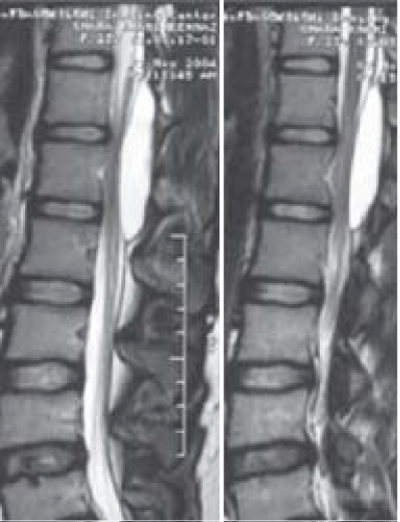
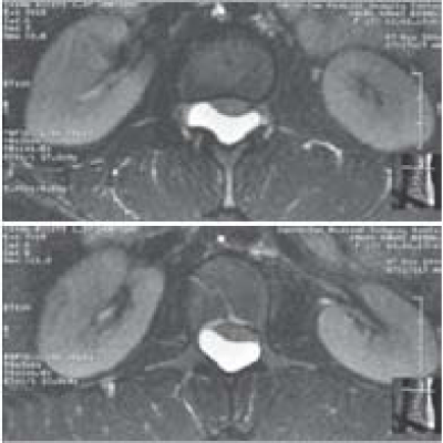
On MRI (Figures 1 and 2), arachnoid cyst was solitary in 12 cases. Only one patient presented 2lumbar arachnoid cysts. The location of the cyst was epidural in 11 cases and intramedullary in 2 cases. All the cysts were posterior or postero lateral. All cysts had cerebrospinal intensity on T1 and T2 weighted imaging.
The topography of the cysts was Thoracic in 8 cases, Lumbar in 4 cases and cervico-thoracic in one case.
12 patients underwent surgery. Only one patient was not surgically managed. It was about an 8 year-old girl that presented a weakness of lower limbs after being treated for scoliosis by a corset. Imaging showed a thoracic epidural arachnoid cyst. But after removal of the corset was noticed a significant improvement of the symptomatology.
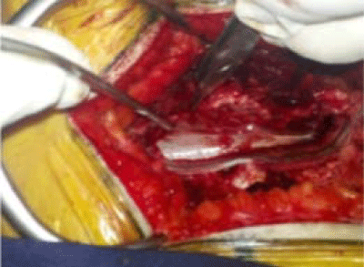
10 patients had total cyst excision after laminectomy (Figure 3). A solitary dural defect was found in 10 patients and could be tightly closed. Multiple dural defects were found in one case and couldn’t be closed. A shunt between the cystic cavity and subarachnoid space was performed for this patient.
One patient had a giant arachnoid cyst enlarging foramina from D3 to D12. The option of lomboperitoneal shunt was preferred for this patient, but no clinical improvement could be obtained by this approach.
Postoperative course was uneventful for all patients except one who had meningitis managed with antibiotics.
The mean follow up was about 17 months. Improvement of initial symptomatology was noticed in 9 cases.
Discussion
Spinal arachnoid cysts are rare lesions that occur inside the spinal canal, representing 1% of all spinal expansive processes [3]. Pathogenesis of these lesions is still subject of debate. Some studies considered intradural arachnoid cysts as sequels of chronic inflammation [1]. A defect of the dura and the herniation of the arachnoid tissue is supposed to be the main mechanism for the development of spinal epidural cysts [4]. A congenital etiology was also proposed by Perret [5]. He supposed arachnoid cysts would be the result of widening of the septum posticum. But none of these theories could make unanimity through neurosurgical committees as the origin of the onset of this defect is still not proved [6].
There is also still no consensus about classification for spinal arachnoid cysts. Nabors [7] proposed a classification in which he divided spinal cysts into three major categories: epidural meningeal cyst without nerve root fibers (type I), epidural meningeal cysts with neural fibers (type II, the so-called Tarlov cysts), and intradural meningeal cysts (type III).
In our series, epiradural cysts were found in 84% of the cases and 16% were intramedullary, but non intradural extramedullary cysts were found.
To the best of our knowledge, this series is one of the largest in the purpose of arachnoid cysts after the ones of Netra [3,8].
This pathology seems to affect women with higher rates than men. Almost 60% of the patients reported in the seriesof Jae and Tokmak were female gendered [2,8].
In our series, 76% of the patients were female gendered, which isconsistent with literature features. In previous studies concerning spinal arachnoid cysts, medical background had not been specified. But we do think that patient history is important to define, mainly when it is about spinal pathologies, even when not surgically managed. In fact, we had in our series two patients who were managed for cyphosoliosis several years before the diagnosis of spinal arachnoid cysts. We can presume that spinal deformities can be incriminated as predisposing factor towards the development of spinal arachnoid cysts. On the other hand, we had also 2 patients followed for sarcoidosis and Neuro fibromatosis.
Multiple arachnoid cysts are rare and mainly reported in the childhood [9]. 12 of our patients had solitary cysts and only 4 years old girl had two extradural lumbar cysts with multiple dural defects discovered peroperative.
Initial symptomatology can be various depending on the level of neurologic compression. Paraparesis is the most reported symptom in the literature. Urinary disorders can also be noticed especially if the sacral roots are affected. Sensitive deficits are rarely reported [9,10]. Weakness of the lower limbs was as well the most frequent in our series with a rate of 69%. Urinary disturbance was found in 53%. Dysesthesia was noticed in 30% of our patients.
MRI is the most accurate method for diagnosis and study of the spinal arachnoid cysts. CT scan can be useful to show indirect signs such as bone erosion or foraminal enlargement [2]. Myelography can precise in some cases the location of a communication between the cyst and the subarachnoid space [11].
Epidural location is the most common localization among spinal arachnoid cysts [12]. On the opposite, intradural extramedullary would bethe rarest. Our finding are concordant with those of the literature, as 84% were epidural, 16% were intramedullary, but no one was intradural extramedullary.
Surgery is consideredas the Gold Standard for treating symptomatic spinal arachnoid cysts [13,14]. The most reported approach has for principles, after laminectomy, the excision of the cyst and the tight closure of the dural breach. This is a classic approach that provided the best results [7]. Other techniques can be discussed in the cases in which excision of the cyst is not possible, or when the dural breach is too wide making tight closure tough. In these cases shunting and cyst punction are reported with more controversial outcomes [1,14].
In our series, 12 patients underwent surgery. 10 patients had total cyst excision after laminectomy. A solitary dural defect was found in 10 patients, and was closed with the help of a plasty. Multiple dural defects were found in one case and couldn’t be closed. A shunt between the cystic cavity and subarachnoid space was performed for that patient. One patient had a giant arachnoid cyst enlarging foramina from D3 to D12. A limbo peritoneal shunt was performed for this patient.
The outcome varies depending on the width of the breach and therapeutic approaches. When a total excision with tight closure of the breach are performed, clinical evolution is satisfying and cyst recurrence is very rare [1,2]. All the patients that were managed through this technique in our series had a good outcome, with total or partial regression of the symptoms. Prognosis is worse when an excision of the cyst is not possible, or when the dural breach is too wide making tight closure tough. In our series, no significant improvement couldbe noticed for the patient who was operated through cyst shunting.
Conclusion
Spinal arachnoid cyst is a rare but curable pathology. Cysts excision and dural defect closure seems to be the most efficient treatment for symptomatic cysts. More conservative approaches like shunt or puncture have worse outcomes and should be reserved for specific cases.
References
- Kumar A, Sakia R, Singh K, Sharma V. Spinal arachnoid cyst. J Clin Neurosci. 2011; 18: 1189-1192.
- Tokmak M, Özek E, Iplikcioglu A. Spinal Extradural Arachnoid Cysts: A Series of 10 Cases. J Neuro Surg A. 2015; 76: 348-352.
- Netra R, Min L, Shao Hui M, Wang J, Bin Y, Ming Z. Spinal extradural meningeal cysts : an MRI evaluation of a case series and literature review. J Spinal Disord Tech. 2011; 24: 132-136.
- Kim K, Chun S, Chung S. A case of symptomatic cervical perineural (Tarlov) cyst : clinical manifestation and management. Skelet Radiol. 2012; 41: 97-101.
- Perret G, Green D, Keller J. Diagnosis and treatment of intradural arachnoid cysts of the thoracic spine. Radiology. 1962; 79: 425-429.
- Lee C, Hyun S, Kim K, Jahng T, Kim H. What is a reasonable surgical procedure for spinal extradural arachnoid cysts : is cyst removal mandatory? Eight consecutive cases and a review of the literature. Acta Neurochir (Wien). 2012; 154: 1219-1227.
- Nabors M, Pait T, Byrd E. Updated assessment and current classification of spinal meningeal cysts. J Neurosurg. 1988; 68: 366-377.
- Oh J, Lee D, Kim T, Yi S, Ha Y, Kim K, et al. Thoracolumbar extradural arachnoid cysts: a study of 14 consecutive cases. Acta Neurochir (Wien). 2012; 154: 341-348.
- Cloward R. Congenital spinal extradural cysts: case report with review of literature. Ann Surg. 1968; 168: 851-864.
- Gortvai P. Extradural cysts of the spinal canal. J Neurol Neurosurg Psychiatry. 1963; 26: 223-230.
- Congia S, Coraddu M, Tronci S, Nurchi G, Fiaschi A. Myelographic and MRI appearances of a thoracic spinal extradural arachnoid cyst of the spine with extra- and intraspinal extension. Neuroradiology. 1992; 34: 444-446.
- Sharma A, Sayal P, Badhe P, Pandey A, Diyora B, Ingale H. Spinal intramedullary arachnoid cyst. Indian J Pediatr. 2004; 71: 65-67.
- Morio Y, Nanjo Y, Nagashima H, Minamizaki T, Teshima R. Sacral cyst managed with cyst-subarachnoid shunt: a technical case report. Spine. 2001; 26: 451-453.
- Bellavia R, King J, Naheedy M, Lewin J. Percutaneous aspiration of an intradural/extradural thoracic arachnoid cyst: use of MR imaging guidance. J Vasc Interv Radiol. 2000; 11: 369-372.
Citation: Bahri K, Chabaane M, Gader G, Bouali S, Jemel H and Zammel I. Spinal Arachnoid Cysts: Clinical and Technical Study. Clin Oncol. 2019; 2(1): 1004.
PDF DownloadClinics Oncology © All Rights Reserved. 2018


