ISSN: 2688-562X
Case Report
Metastasis of Esophageal Squamous Cell Carcinoma to Gastrotomy Site: A Case Report
Mei Kang and Yichun Wang*
- Department of Radiation Oncology, the First Affiliated Hospital of Anhui Medical University, China
- *Corresponding author: Yichun Wang, Department of Radiation Oncology, the First Affiliated Hospital of Anhui Medical University, Hefei, China;
- Received: Jul 11, 2019 Accepted: Jul 23, 2019 Published: Jul 25, 2019
Abstract
A 67-year-old man with abdominal distension underwent gastrointestinal fiberscopy (GIF) and was found to have a lesion with in the lower esophagus and cardia. The patient received the endoscopic submucosal dissection (ESD) for the esophagus carcinoma and have the resection of gastric lesions. Pathological examination of both tumors demonstrated moderately differentiated squamous cell carcinoma. A total of 60Gy in 30 fractions of three-dimensional conformal radiation therapy (3D-CRT) was administered to the esophageal carcinoma. No local recurrence was observed in either the esophagus or the stomach during follow-up. Considering the dismal prognosis of esophageal carcinoma patients with intramural metastasis to the stomach, a watchful follow-up is needed.
Keywords:
Esophageal carcinoma; Gastric metastasis; Radiation therapy
Introduction
The presence of metastasis in the stomach from esophageal squamous cell carcinoma is relatively rare, lack of related studies in the domestic and foreign [1-3]. It have been proved that the prognosis of esophageal cancer with gastric metastasis is poor, and the 5-year survival rate is 15%-25% [4,5]. Herein we report a case of esophageal squamous cell carcinoma with gastric wall and left gastric lymph node metastasis, and investigate the metastasis mechanism, treatment and prognosis
Case Report
A 67-year-old man complained of intermittent upper abdominal distension. Gastroscopy showed that high-grade intraepithelial neoplasia of esophageal squamous growth 29cm from the incisors (Figure A), He underwent the ESD for the esophagus lesion (Figure B), pathologic proved that squamous cell carcinoma invaded the submucosa with a depth of more than 750 um. Computed tomogram (CT) of Chest and abdomen showed thickening of the middle esophageal wall, considering esophageal cancer, lesions between the stomach at the lower esophagus and cardia, and the possibility of stromal tumors or leiomyomas (Figure C, D). Barium meal of upper digestive tract asserted equally the rigid and filling defect of middle thoracic esophageal tube wall was observed, and no obvious niche and filling defect was found in gastric wall (Figure E). Because of the attack of cerebral infarction, the patient rejected the surgery and refuse to the chemotherapy in the followup planning. He underwent radiation therapy only for the primary esophagus. A 3D-CRT plan was designed, the planning target volume (PTV) was 60Gy in 30 fractions. After the recovery of the radiotherapy, and patients received the resection of partial gastrectomy plus small curvature lymphadenectomy. The operation showing that the tumors were located on the lesser curved side of the stomach, with a diameter of about 3cm, and there was a swollen lymph node with a diameter of about 2.5cm. Postoperative pathology showed moderately differentiated squamous cell carcinoma, local cancer tissue adjacent to the incision margin, considered as esophageal cancer proliferation, the left gastric lymph node also showed moderately differentiated squamous cell carcinoma. A follow-up study was performed 2 months after the surgery, no recurrence and metastasis was found.
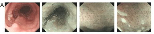
Figure A: Gastroscopy findings: A slightly rough mucosa, reddish surface, irregular margin, the widest circumference was 2/3 weeks 29-34 cm away from the incisor. NBI + magnifying gastroscopy showed that the lesion was brown, IPCL was type B1, local type B2.
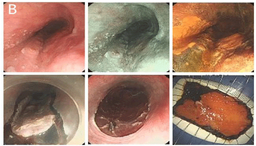
Figure B: From 26 to 34 cm from the incisor, the mucosa was slightly rough, the surface was reddish and the edge was irregular, irregular light or non-staining areas were observed after iodine staining. Normal saline, sodium hyaluronate and kidney were injected under the mucosa. ESD was performed after adenine elevation, and the lesion was completely dissected.
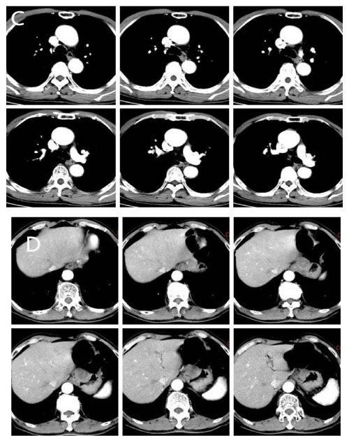
Figure C, D: CT of Chest and abdomen: The middle esophagus was obviously thicker, and the lesions of the stomach with the largest diameter of about 3 cm were found in the lower esophagus and cardia, showed uneven density enhancement.
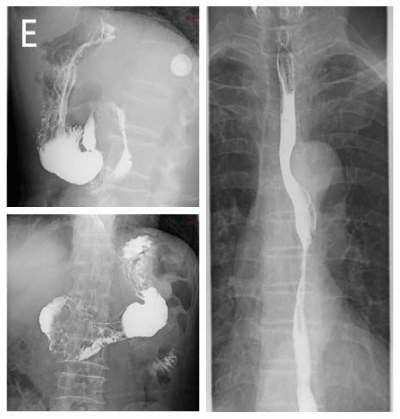
Figure E: From 26 to 34 cm from the incisor, the mucosa was slightly rough, the surface was reddish and the edge was irregular, irregular light or nonstaining areas were observed after iodine staining. Normal saline, sodium hyaluronate and kidney were injected under the mucosa. ESD was performed
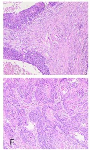
Figure F: From 26 to 34 cm from the incisor, the mucosa was slightly rough, the surface was reddish and the edge was irregular, irregular light or non-staining areas were observed after iodine staining. Normal saline, sodium hyaluronate and kidney were injected under the mucosa. ESD was performed after adenine elevation, and the lesion was completely dissected.
Discussion
Esophagus has a complex lymphatic drainage system. Its submucosa and lamina propria have a longitudinal lymphatic network and a long drainage area, which can be directly drained to mediastinal lymph node or thoracic duct [6].Therefore, no matter which segment of esophageal carcinoma, or even early esophageal carcinoma, the range of lymph node metastasis can be from neck to abdominal, it showed the pattern of ”jumping metastasis”. Saito proposed the diagnostic criteria for metastasis of esophageal carcinoma to gastrotomy in 1985 [4]: 1) The histological diagnosis of the two lesions was the same regardless of the degree of cell differentiation; 2) the lesions were discontinuous without considering the invasion of blood vessels or lymphatics; 3) the lesions in the stomach were discontinuous with the metaplasia of squamous epithelium or squamous epithelium in the stomach. In this case, the gastric metastases were located in the small curvature of the stomach. The diameter of the lesion was about 3cm, and the lymph node with a diameter of 2.5cm were found in the distal part of the small curvature. The pathological results were consistent with the pathological results of the esophageal lesions, It is suggested that the diagnosis of metastasis of esophageal and gastric cancer is consistent with that of metastasis of esophageal and gastric carcinoma. Metastasis of esophageal squamous cell carcinoma to stomach are often found unexpectedly in autopsy or examination. The incidence of esophageal cancer metastases in autopsy is about 0-15% [1]. At present, there is no systematic regression study, most are the case reports.
The mechanism of gastric metastasis of esophageal carcinoma has not been determined. At present, most scholars believe that the possible mechanism of metastasis is that tumor cells may metastasize to the stomach through the submucosal lymphatic system. Gastric metastasis of esophageal cancer may be correlated with its anatomical structure. The esophageal wall and gastric wall belong to the digestive system, so their structures are similar. They tube wall are divided into six sections: epithelium, lamina propria, muscular layer of mucosa, connective tissue of submucosa, muscular layer and serosa. The esophageal wall has no adventitia and is serosa layer. Vessels existing in all layers except epithelium and its basement membrane. They run mainly longitudinally in the lamina propria and the submucosa and circumferentially in the intermuscular space. Vessels in lamina propria and muscular is mucosae connect to submucosa. Vessels in submucosa are generally located in the outer margin of this layer. They connect to thoracic duct, regional lymph node, and possibly to intermuscular lymphatic plexus and paraesophageal node. This complex anatomical structure and abundant lymphatic vessels and vascular communicating branches may be the anatomical basis of gastric metastasis of esophageal cancer. Zhu [6], studied 11 patients with esophageal squamous cell carcinoma which lesions located in the middle and lower section, while the metastases in the stomach located in the upper 1/3 of the stomach, the depth of invasion of the primary lesions was exceeded to the submucosa. Four of them were in stage T1-T2 and seven in T3-T4. Therefore, it is likely to be through blood vessels or lymphatic vessels to transfer to the gastric wall of esophagus carcinoma. Simultaneously, some scholars have put forward the idea that esophageal carcinoma implantation metastases to the stomach. Hassan Hameed [7], reported the first case of gastric metastasis after endoscopic gastrostomy in patients with esophageal cancer due to dysphagia, suggesting that the tumor cells could directly fall off to the gastric wall by some way leading to implantation metastasis. Satoshi Asai [8], also provided a case of esophageal squamous cell carcinoma complicated with gastric adenocarcinoma. submucosal tumors appeared in the scar area after ESD, pathological findings performed that was moderately differentiated squamous cell carcinoma, and no other lesions suggested blood metastasis.
Gastric metastasis of esophageal carcinoma is considered to be a poor factor, because of the low incidence of gastric metastasis of esophageal cancer and insufficient attention in clinic, gastroscopy cannot inspect the gastric due to the narrowing of the esophageal lumen, and if the lesions in the stomach are small, imaging examination is easy to miss diagnosis, sometimes misdiagnosed as gastrointestinal stromal tumors or primary tumors of the stomach, so the prognosis is poor. Several studies [4], shown that the cumulative survival rate of gastric metastasis of esophageal cancer is significantly lower than that of non-gastric metastasis of esophagus. The median survival time of patients with gastric metastasis of esophageal cancer after operation is 5.8 months, and the 3-year survival rate is about 9%. The survival rate of patients with gastric metastasis is not significantly improved even received preoperative chemotherapy. Therefore, if esophageal carcinoma merged gastric lesions, we need to be vigilant about the possibility of gastric metastasis, and need to combine multiple examinations to improve the detection in order to make early diagnosis and earlier treatment.
There is no uniform standard for the treatment of gastric metastasis from esophageal carcinoma, nor relevant guidelines or consensus recommendations. Most patients are treated according to the routine diagnosis and the experience of clinicians of the esophagus cancer. Surgery is recommended for without distant metastases, but the prognosis is still poor. At present, surgery is the main treatment in clinic. Because most of the gastric metastases are located in the cardia and small curvature, which can be resected by surgery and reconstruction of digestive tract by esophagogastrostomy. Naoko Hashimoto [9], reported a patient with gastric metastasis of esophageal cancer (cT3N0M1b) by combination of breath-gating technique and 3D-radiotherapy, the total dose was 60 Gy/30f after radiotherapy, and only grade 1 mucositis occurred, no recurrence or metastasis in the follow-up 17 months after radiotherapy. Therefore, radiotherapy can also be used as one of the therapy for esophageal cancer with gastric metastasis. In this case, esophageal cancer has undergone radical radiotherapy, and gastric lesions have been resected surgically. No recurrence or metastasis of the tumors has been observed.
Conclusion
Gastric metastasis of esophageal carcinoma is rare, the mechanism of metastasis is uncertain, it may be metastasized by lymphatic vessels or implantation, and related to the anatomical structure of esophageal and gastric wall. The overall prognosis is poor, the combination of different tests helps provide detection rate. Surgery or radiotherapy can be used the effective treatment measures.
References
- Yuma Ebihara, Masao Hosokawa, Satoshi Kondo, Hiroyukikatoh. Thirteen cases with intramural metastasis to the stomach in 1259 patients with oesophageal squamous cell carcinoma. Eur J Cardiothorac Surg. 2004; 26: 1223-1225.
- Takubo K, Sasajima K, Yamashita K, Tanaka Y, Fujita K. Prognostic significance of intramural metastasis in patients with esophageal carcinoma. Cancer. 1990; 65: 1816-1819.
- Kato H, Tachimori Y, Watanabe H, Itabashi M, Hirota T, Yamaguchi H, et al. Intramural metastasis of thoracic esophageal carcinoma. Int J Cancer. 1992; 50: 49-52.
- Saito T, Iizuka T, Kato H, Watanabe H. Esophageal carcinoma metastatic to the stomach: a clinicopathological study of 35 cases. Cancer. 1985; 56: 2235-2241.
- Zhang SW, Zheng RS, Zuo TT,Zeng HM, Chen WQ, HE J. Mortality and Survival Analysis of Esophageal Cancer in China. Chinese J Oncol. 2016; 38: 709-715.
- Zhu YJ, Xue LY, Li Y, J, iang J, Liu TT, Jiang LM, et al. Intramural gastric metastasis of esophageal squamous cell carcinoma: an analysis of imaging findings and clinicopathological features. Chinese J Oncol. 2017; 39: 509- 513.
- Hassen Hameed, Yakub Iqbal Khan. Metastsis of carcinosarcoma of oesphagus to gastrotomy site. J Oral Maxillofacial Surg. 2009: 643-644.
- Satoshi Asai, Koutarou Takeshita, Yuki Kano. Implantation of esophageal cancer onto post-dissection ulcer after gastric endoscopic submucosal dissection.WJG. 2015; 22: 2855-2860.
- Naoko Hashimoto, Jin Iwazawa, Hisashi Abe. Radiation therapy for gastric wall metastasis from esophageal carcinoma. Jpn J Radiol. 2010; 28: 227- 230.
Citation: Yichun W. Metastasis of Esophageal Squamous Cell Carcinoma to Gastrotomy Site: A Case Report. Clin Oncol. 2019; 2(2): 1011.
PDF DownloadClinics Oncology © All Rights Reserved. 2018
